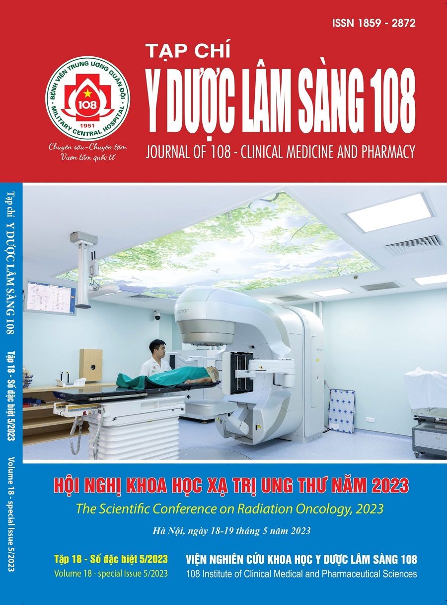
Evaluation of the contralateral breast dose in primary breast cancer radiotherapy using intensity modulated radiation therapy technique and field in field technique
Main Article Content
Keywords
Abstract
Objective: To evaluate the dose received by the contralateral breast in radiation therapy of primary breast cancer using IMRT technique compared to FiF technique. Subject and method: There were 32 CT series of patients in this study, divided into Group G1: Nodes including Internal Mammare Node (IMN) and Group G2: nodes without IMN. For each patient, two different plans were created for the entire treated breast (including IMRT and FiF techniques) with only one CT simulator series. The prescription is 5000cGy per 25 fractions. The dosimetric parameters of the planning target volume for dose evaluation and the organs at risk for each planning technique were compared. Dose to the contralateral breast was compared especially between two techniques with Dmean, Dmax, D100cGy, D500cGy, and dose at the center and at 4cm medial position the center of the contralateral breast. Result: Doses to PTV and organs at risk by both techniques meet the criteria for evaluation of the plan. The Dmax of the contralateral breast using IMRT and FiF techniques were 839 ± 98cGy and 3537 ± 152cGy respectively in group G1, while they were 543 ± 124cGy and 1035 ± 85cGy respectively in group G2. The dose at the center of the contralateral breast and at 4cm position medial the center of the contralateral breast using the IMRT technique was reduced by 2 times compared to that using the FiF technique when comparing both groups. Evaluations of 500cGy and 300cGy coverage of the contralateral breast using IMRT and FiF techniques showed no difference. The 100cGy dose covering the contralateral breast using IMRT in group G1 was 89.35 ± 0.87%, which was higher than that using the FIF technique was 69.41 ± 1.24%, while the figures for group G2 were 70.58 ± 2.02% and 42.44 ± 1.53%, respectively. The average Dmean of the contralateral breast obtained using the IMRT technique was 393 ± 137cGy, which was higher than that using the FiF technique of 273 ± 95cGy (group G1); IMRT 305 ± 201cGy was higher than FiF 206 ± 102cGy (group G2). Conclusion: Compared with FiF technique, IMRT used in breast cancer radiotherapy resulted in the reduction of the high level dose and maximum dose to the contralateral breast. FiF technique has better control in low-level dose, Dmean to the contralateral breast than IMRT technique.
Article Details
References
2. Yadav BS, Sharma SC, Patel FD, Ghoshal S, Kapoor RK (2008) Second primary in the contralateral breast after treatment of breast cancer. Radiother Oncol 86(2): 171-176.
3. Krueger EA, Fraass BA, Pierce LJ (2002) Clinical aspects of intensity-modulated radiotherapy in the treatment of breast cancer. Semin Radiat Oncol 12(3): 250-259. doi: 10.1053/srao.2002.32468.
4. Hall EJ, Wilkins LW (1994) Radiobiology for the Radiologist. 5th edition: 144-165.
5. Health NIo, Statement CDC (1991) Treatment of Early-Stage Breast Cancer. NIH Consens Statement. JAMA 265: 391-395.
6. Hidekazu Tanaka MI, Takahiro Yamaguchi, Kae Hachiya, Takahiko Yajima, Masashi Kitahara, Katsuya Matsuyama, Satoshi Goshima, Manabu Futamura, and Masayuki Matsuo (2017) High Tangent Radiation Therapy With Field-in-Field Technique for Breast Cancer. Breast Cancer (Auckl); 11: 1-5.
7. https://moh.gov.vn/hoat-dong-cua-dia-phuong/-/asset_publisher/gHbla8vOQDuS/content/tinh-hinh-ung-thu-tai-viet-nam.
8. International Agency for Research on Cancer; GLOBOCAN 2020.
9. Boice JD, Blettner M, Stovall M, Flannery JT (1992) Cancer in the contralateral breast after radiotherapy for breast cancer. N Engl J Med 86(2):171-6;
10. Loïc Feuvret Md, Georges Noël, M.D., Jean-Jacques Mazeron, M.D., Ph.D, and Pierre Bey Md (2006). Conformity Index: A Review; 64(2):333-42;
11. RTOG 1304 (2016) R. A Randomized phase III clinical trial evaluating post-mastectomy chestwall and regional nodal xrt and post-lumpectomy regional nodal XRT in patients with positive axillarynodes before neoadjuvant chemotherapy who convert to pathologically negative axillary nodes after neoadjuvant chemotherapy.
12. Zakiya Salem Al-Rahbi ZAM, Ramamoorthy Ravichandran, Fatma Al-Kindi, Cheriyathmanjiyil Anthony Davis, Saju Bhasi, Namrata Satyapal, and Balakrishnan Rajan (2013) Dosimetric comparison of intensity modulated radiotherapy isocentric field plans and field in field (FIF) forward plans in the treatment of breast cancer. Journal of Medical Physics: 22-29.
13. Zeverino M, Petersson K, Kyroudi A, Jeanneret-Sozzi W, Bourhis J, Bochud F, Moeckli R (2018) A treatment planning comparison of contemporary photon-based radiation techniques for breast cancer. Phys Imaging Radiat Oncol 7:32-38. doi: 10.1016/j.phro.2018.08.002.
 ISSN: 1859 - 2872
